
DiverInterCom KCore Scuba · Decompression Science & Practice
Every Dive Is a Decompression Dive
Every scuba dive involves decompression, even when no mandatory decompression stops are required.
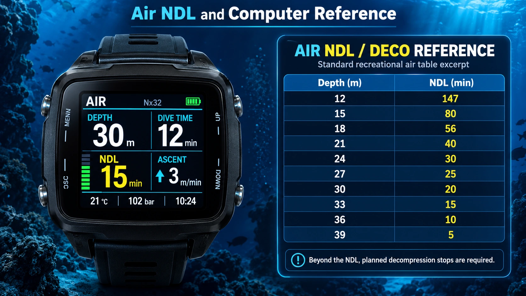
For recreational divers, one of the most important concepts is the No-Decompression Limit (NDL). It represents how long a diver can remain at a given depth before an ascent following the selected model's rules would require mandatory decompression stops.
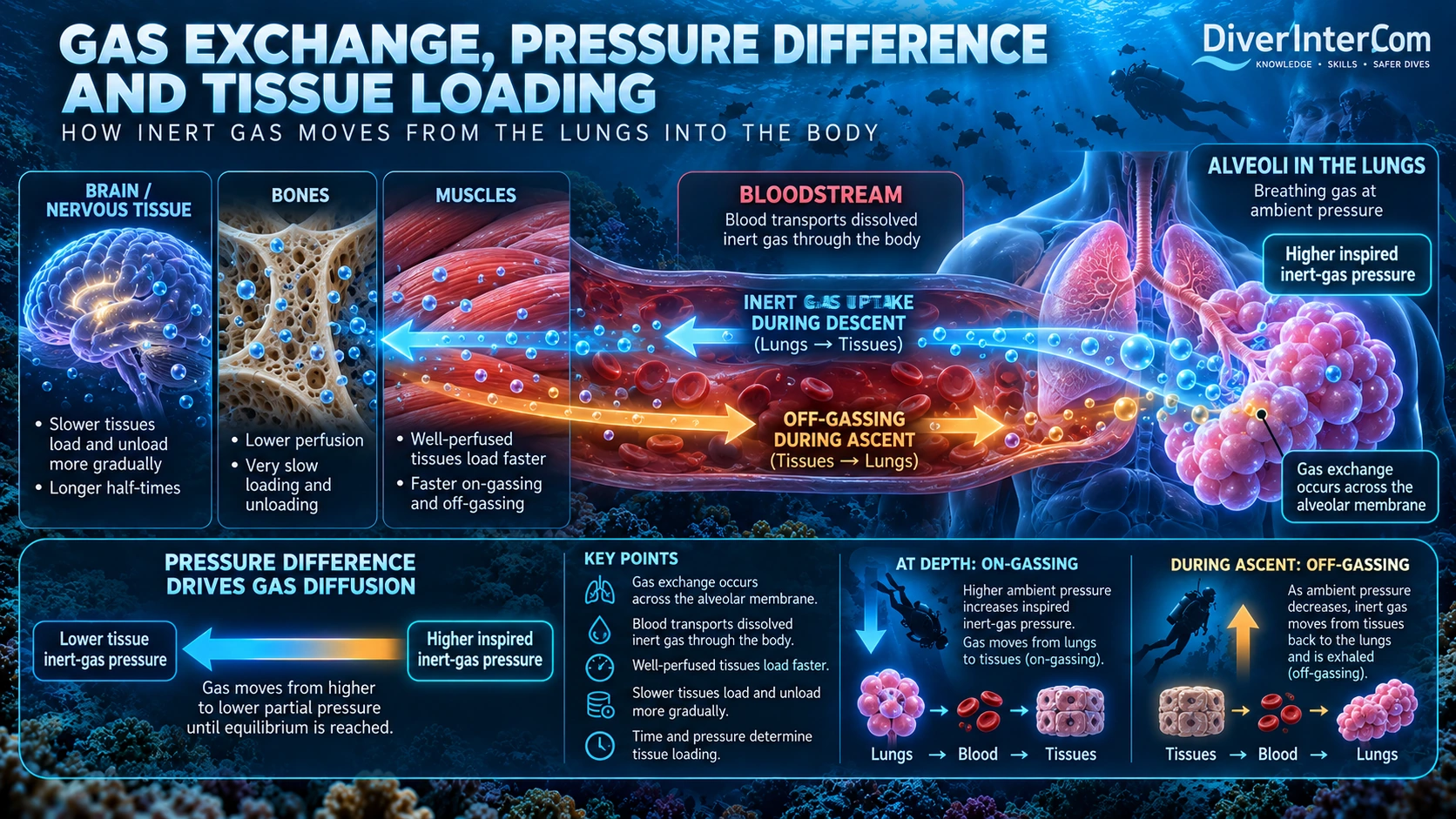
While breathing compressed air, the body absorbs inert gas, primarily nitrogen. This is called on-gassing. As the diver ascends and surrounding pressure decreases, dissolved nitrogen begins to leave the tissues through off-gassing.
During a dive within the NDL, the model estimates that tissue loading permits a controlled ascent without mandatory decompression stops. Gas elimination is not instantaneous: different tissues exchange gas at different rates, and off-gassing continues after surfacing.
A slow, controlled ascent is essential. A common recreational guideline is no faster than 9 metres per minute, while always following the specific dive computer's or training agency's ascent-rate instructions.
Even on no-stop dives, a safety stop of about 3 minutes at 3–5 metres is commonly recommended when conditions permit. It provides additional time for inert-gas elimination and may reduce decompression stress.
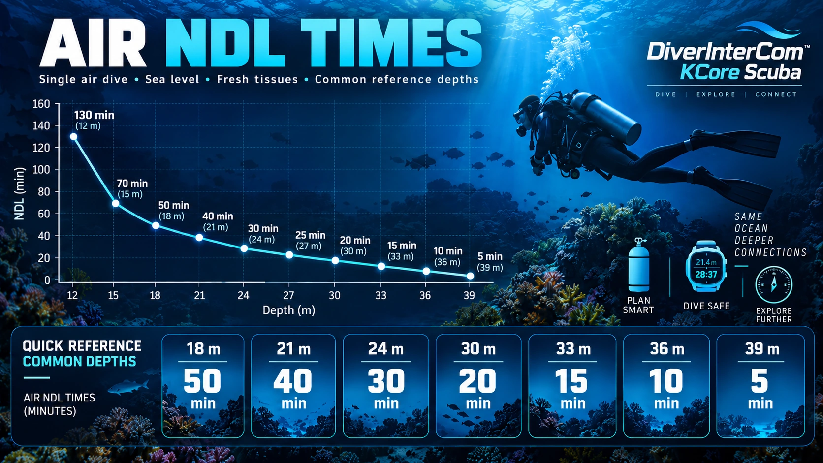
The Air NDL chart above illustrates single-dive table limits for air at sea level with no residual loading. A table is not a universal physiological threshold. Use the correct full table, training procedures, dive plan and properly configured computer for actual dives; account for repetitive dives and conditions.
What is behind the numbers?
Understanding Decompression Models
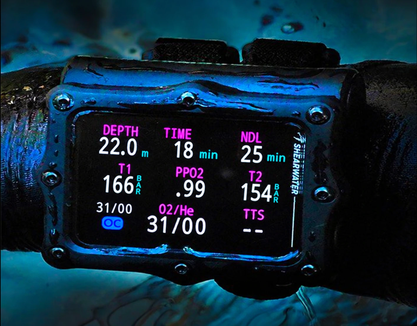
Modern dive computers use mathematical models to estimate how inert gases such as nitrogen and helium enter and leave the diver's tissues. These calculations help determine no-decompression limits, decompression ceilings and required ascent stops.
Bühlmann ZH-L16 is one of the most widely used decompression model families in modern dive computers. It represents the diver with 16 theoretical tissue compartments that exchange inert gas at different rates. Many computers combine Bühlmann with Gradient Factors (GF), settings that modify the supersaturation allowed by the model.
RGBM (Reduced Gradient Bubble Model) and VPM (Varying Permeability Model) are other influential approaches incorporating assumptions about microscopic gas bubbles and their behavior during decompression. RGBM has appeared in recreational dive computers, while VPM has been used in technical decompression-planning software. Specific manufacturer implementations may differ.
Different models, configurations and implementations can produce different NDLs or decompression schedules for the same exposure. A shorter NDL or deeper decompression stops do not necessarily mean a safer dive.
For recreational divers, the first priority is understanding the displayed NDL, following ascent guidance and allowing adequate margins. As diving becomes more advanced, tissue compartments, gradient factors and bubble-model assumptions become increasingly relevant.
Conservatism and Gradient Factors
Many modern dive computers allow the diver to increase or decrease the conservatism of the decompression calculation. In practical terms, a more conservative setting usually means a shorter NDL, and on decompression dives it may require longer or earlier stops.
With Bühlmann-based computers, this is often adjusted using Gradient Factors (GF). GF Low mainly influences the deeper part of a decompression ascent and where the first stop may appear. GF High has a strong influence on the shallower part of the ascent and on the no-decompression limit.
A lower GF setting generally makes the computer more restrictive, while a higher GF setting allows more of the model’s calculated supersaturation. However, different computers may still give different results even when the same GF numbers are selected.
Important: A shorter NDL or a longer decompression schedule does not automatically prove that a dive is safer. These settings are model-based tools, not direct measurements of the diver’s body.
Conservatism, Safety Margins and Gradient Factors
A more conservative Gradient Factor setting provides a larger calculated safety margin between the diver's modeled tissue-gas pressure and the Bühlmann M-value limits.
Lower GF High values generally provide a greater margin by allowing less supersaturation, resulting in shorter NDLs and potentially longer decompression requirements.
Higher GF High values are less conservative. They allow greater supersaturation, providing a smaller calculated safety margin but often longer NDLs and shorter decompression obligations.
In general, reducing decompression stress through appropriate conservatism is intended to lower the likelihood of decompression sickness (DCS), commonly known as the bends.
However, a larger mathematical safety margin does not guarantee freedom from DCS. Lowering GF Low can introduce deeper stops, which are not necessarily safer. Individual physiology, dive conditions and the complete decompression profile also influence risk.

Inside the decompression model
The 16 Tissues, M-Values and Gas Loading
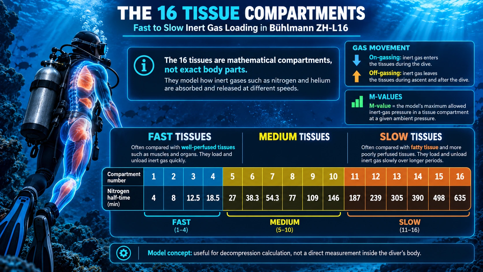
Bühlmann ZH-L16 represents the diver's body using 16 theoretical tissue compartments. These are not 16 exact real body parts, but mathematical compartments used to model how inert gases such as nitrogen and helium enter and leave the body at different rates.
Some compartments behave like fast tissues. In practical teaching terms, these are often compared to well-perfused tissues such as muscles and other body tissues with relatively good blood supply. They can take up and release inert gas more quickly.
Other compartments behave like slow tissues. These are often compared with fatty tissue and more poorly perfused tissues, where inert gas loading and unloading happens more slowly over a longer time. This is why not every part of the body reacts to depth changes at the same speed.
During the dive, inert gas enters the body through on-gassing. During ascent and after surfacing, excess inert gas leaves the tissues through off-gassing. The balance between these two processes is one of the core ideas behind decompression theory.
An M-value is the maximum inert-gas pressure that the model allows in a tissue compartment at a given ambient pressure. It is a modeled upper limit of tolerated supersaturation. Dive computers use these limits to estimate no-decompression limits, decompression ceilings and ascent safety margins.
From no-stop to mandatory stops
When an NDL Dive Becomes a Decompression Dive
During a normal recreational dive, the diver remains within the No-Decompression Limit (NDL). This means the diver can ascend directly to the surface at a controlled ascent rate without requiring mandatory decompression stops, according to the dive computer's decompression model.
As the diver spends more time at depth, the body continues absorbing inert gases, primarily nitrogen when breathing air. This process is called on-gassing. The deeper and longer the exposure, the greater the inert-gas loading in different body tissues.
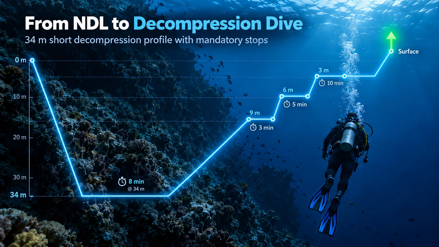
When the diver exceeds the calculated NDL, the dive enters mandatory decompression. The computer now calculates that a normal direct ascent would exceed its permitted supersaturation limits. The diver must follow a controlled ascent with the required decompression stops before surfacing.
During ascent, the surrounding water pressure gradually decreases. As ambient pressure falls, dissolved inert gas leaves the tissues, moves into the blood, is transported to the lungs, and is then removed from the body during exhalation. This process is called off-gassing.
The purpose of staged decompression is to allow enough time for safe inert-gas elimination while controlling supersaturation and limiting the conditions that promote harmful bubble formation. The dive computer continuously recalculates the diver's decompression obligation and may display a ceiling, required decompression stops, and time to surface (TTS).
If a diver ascends too quickly after exceeding the NDL, excessive supersaturation may occur. Under these conditions, bubbles can form or grow in tissues and blood, potentially causing Decompression Sickness (DCS), commonly called the bends.
Air, nitrox, deco gas and helium
Breathing Gases and Decompression
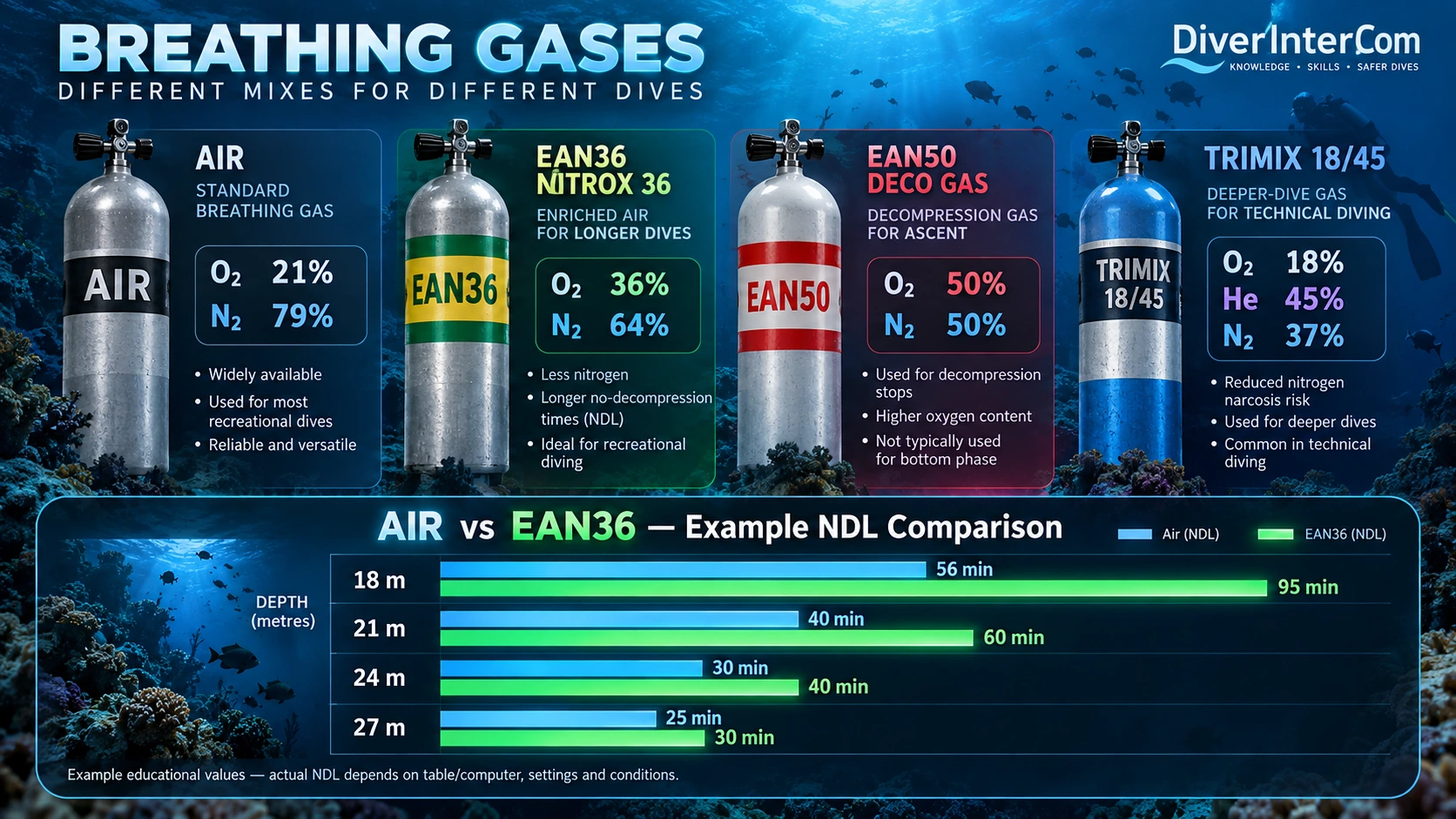
For most recreational scuba divers, compressed air is the standard breathing gas. Air contains about 21% oxygen and 79% nitrogen, and its nitrogen content plays an important role in determining no-decompression limits and decompression requirements.
Nitrox, also called EAN or SafeAir, contains more oxygen and less nitrogen than ordinary air. A common example is EAN36. Because nitrox contains less nitrogen, the diver generally absorbs less nitrogen at a given depth, which can allow a longer NDL compared with air. However, the higher oxygen content also makes the maximum operating depth (MOD) shallower.
A second common use of different gases is the gas switch during decompression. The diver uses a suitable bottom gas for the main part of the dive, then switches during ascent to an oxygen-richer decompression gas, often carried in an additional stage cylinder. Less inert gas and more oxygen can increase the gradient for inert-gas elimination and help reduce decompression stop time.
A third important gas family introduces helium for deeper diving. In gases such as trimix or heliox, nitrogen and oxygen fractions are reduced and helium is added because it is lighter and much less narcotic than nitrogen. This helps reduce narcosis and control oxygen exposure on deeper dives. Helium is still an inert gas and must also be included in decompression calculations.
If a dive computer supports multigas diving, it can calculate decompression in real time using the active gas and programmed backup or decompression gases. It may show the required stops, the ceiling and the total time to surface (TTS). But this only works correctly if the diver programs the right gas or gases into the computer before the dive and confirms any gas switches correctly during the dive.
Real-time decompression insight
Understanding and Using GF99 and Surface GF
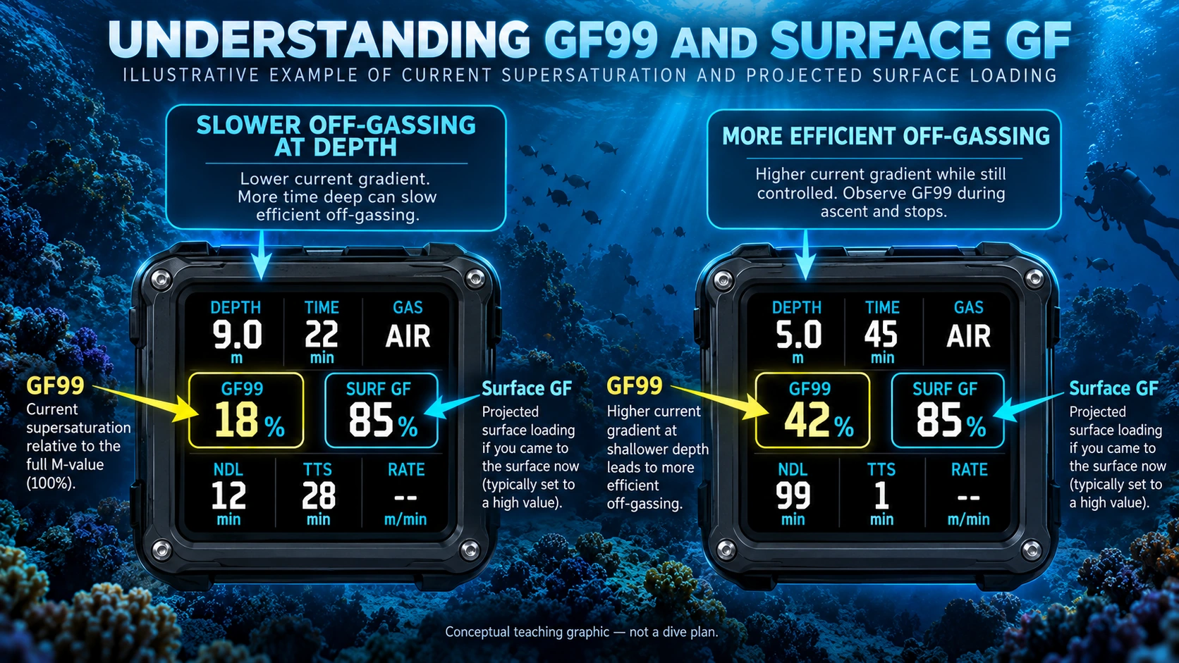
Modern Bühlmann-based dive computers may show two additional values that help divers better understand their decompression status in real time: GF99 and Surface GF (SurfGF).
GF99 shows the current supersaturation of the leading tissue compartment, expressed as a percentage of the full unmodified Bühlmann M-value gradient at the diver’s present depth. In simple terms, it helps show how much of the model’s allowed supersaturation is currently being used.
Surface GF shows what the leading compartment’s gradient factor would be if the diver were taken directly to the surface right now, without giving extra credit for additional off-gassing during the rest of the ascent.
In practical use, divers can observe how GF99 changes during ascent and stops. A lower GF99 while staying deeper may mean a lower current gradient, but it can also mean slower off-gassing. A somewhat higher but still controlled GF99 at a shallower stop may indicate more efficient inert-gas elimination. Surface GF helps show the projected surface loading compared with the selected conservatism setting.
This idea has been discussed clearly by Andy Davis, including in connection with DiveTalk. His teaching encourages divers to understand what the computer is showing instead of only following numbers blindly, especially when observing ascent behaviour and shallow stop efficiency.
From the bends to modern dive computers
A Brief History of Decompression Science
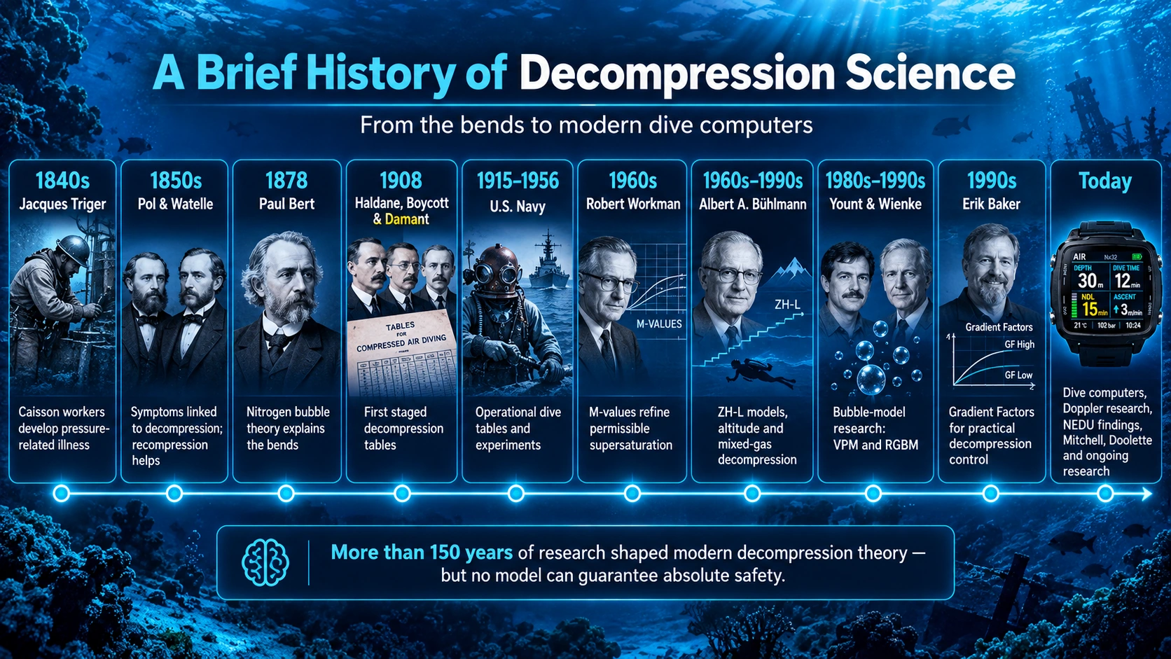
Decompression sickness (DCS), commonly called the bends, was first recognized long before modern scuba diving. In the nineteenth century, workers in pressurized caissons and tunnels developed pain, paralysis and sometimes death after returning too quickly to normal pressure.
Important early names include Jacques Triger, who documented pressure-related illness in caisson workers, and Pol and Watelle, who linked symptoms to decompression and observed that recompression could help.
A major scientific breakthrough came in 1878, when Paul Bert showed that nitrogen dissolved under pressure could form bubbles during decompression. His work provided the scientific explanation for the bends.
In 1908, John Scott Haldane, together with Arthur Boycott and Guybon Damant, developed the first staged decompression tables using the idea of tissue compartments with different gas-loading speeds.
Later, the U.S. Navy advanced decompression research through experiments and dive tables. In the 1960s, Robert Workman introduced the idea of M-values, helping define acceptable supersaturation limits.
Swiss researcher Albert A. Bühlmann further developed decompression theory with the ZH-L model family, which became one of the main foundations of many modern dive computers. Later, David Yount and Bruce Wienke contributed to bubble-model research such as VPM and RGBM, while Erik Baker introduced the practical use of Gradient Factors.
Modern decompression science continues through dive-computer development, Doppler research, military and civilian testing, and the work of experts such as Simon Mitchell and David Doolette. Even today, decompression research is still evolving.
Physiology, physics and decompression models
Understanding Decompression Theory — The Science Behind the Models
Decompression theory is based on a fundamental question: How can a diver return safely from increased underwater pressure to the surface while eliminating the excess inert gases absorbed during the dive?
To understand this, we must look at the interaction between pressure, breathing gases, blood circulation, body tissues and the mathematical models used by our dive computers.
1. The Physiology — How Gases Enter Our Body
When a diver breathes compressed gas underwater, the lungs receive that gas at approximately the surrounding ambient pressure.
Inside the lungs, gas exchange takes place across the extremely thin alveolar-capillary membrane. Oxygen passes into the blood, while carbon dioxide moves in the opposite direction and is exhaled.
Inert gases such as nitrogen and helium also move between the lungs and blood according to their partial-pressure gradients.
The bloodstream transports dissolved inert gases throughout the body. They diffuse from the blood into the fluid surrounding cells and into the cells themselves.
Different tissues absorb inert gases at different rates, depending on blood supply, gas solubility and tissue characteristics.
2. The Physics — Pressure and Gas Laws
Several gas laws explain the behavior of gases during diving.
Boyle's Law: Gas volume decreases as surrounding pressure increases and expands as pressure decreases. This is especially important for gas-filled spaces and the expansion of existing bubbles during ascent.
Dalton's Law: The total pressure of a gas mixture equals the sum of its individual gas partial pressures. Increasing depth increases the partial pressures of the gases we breathe.
Henry's Law: At equilibrium, the amount of gas dissolved in a liquid is proportional to its partial pressure above that liquid, at a given temperature.
Henry's Law is especially important for decompression theory because it explains why more inert gas can dissolve in blood and tissues at increased pressure.
Together, these laws explain why deeper and longer dives can lead to greater inert-gas loading.
3. On-Gassing — Tissue Loading and Saturation
During descent, the partial pressure of inspired nitrogen or helium increases. This creates a pressure gradient between the gas carried by the blood and the gas already dissolved in body tissues.
Inert gas moves along this gradient into the tissues. This is called on-gassing.
The longer the diver remains at depth, the closer the tissue gas tensions approach equilibrium with the inspired inert-gas pressure. This state is called saturation.
However, not all tissues saturate equally quickly. Modern Bühlmann models use 16 theoretical compartments, each with its own gas-exchange half-time. After one half-time at a constant inspired inert-gas pressure, a compartment completes approximately half of its remaining adjustment toward equilibrium.
Fast compartments react quickly; slow compartments may require many hours to approach saturation.
4. Supersaturation — What Happens During Ascent?
During ascent, ambient pressure decreases, reducing the partial pressure of inert gas being breathed.
The tissues may now contain more dissolved inert gas than would be present at equilibrium with the lower inspired inert-gas pressure. This condition is called supersaturation.
Controlled supersaturation is a normal and necessary part of decompression. Without a pressure gradient, excess dissolved inert gas would not be eliminated efficiently.
The critical question is how much supersaturation can be tolerated without creating an unacceptable risk of harmful bubble formation and decompression sickness?
5. Off-Gassing — How Inert Gas Leaves the Body
As the diver ascends, inert gas generally moves from the tissues into the blood, travels through the circulatory system and reaches the lungs. There, it diffuses into the alveolar gas and is removed through exhalation. This is called off-gassing.
The rate of elimination depends on the difference between tissue and inspired inert-gas partial pressures, tissue gas-exchange rates, breathing-gas composition, depth and ascent profile, tissue perfusion and physiological conditions.
Off-gassing can occur at depth, during decompression stops and after surfacing.
6. M-Values — The Mathematical Limits
In dissolved-gas decompression models, each theoretical tissue compartment has a calculated limit for tolerated inert-gas supersaturation at a given ambient pressure. These limits are called M-values.
An M-value represents the maximum modeled inert-gas pressure permitted in a compartment for the surrounding pressure. Exceeding these limits indicates that the decompression schedule has gone beyond the model's accepted boundaries.
However, M-values are mathematical limits, not exact biological thresholds between safety and decompression sickness.
Modern Bühlmann computers often use Gradient Factors to reduce the permitted supersaturation below the original M-value limits, introducing an additional calculated safety margin.
7. Microbubbles and Decompression Sickness
As surrounding pressure decreases, dissolved inert gas can contribute to the formation or growth of microscopic gas bubbles. Small bubbles may occur even after dives without symptoms.
However, excessive bubble formation or growth can interfere with circulation, injure tissues, activate inflammatory processes and contribute to Decompression Sickness (DCS).
Symptoms may include joint pain, skin changes, dizziness, neurological problems, weakness or paralysis. Severe cases can be life-threatening.
Bubble behavior is complex. It depends on more than dissolved-gas pressure alone, which is one reason researchers have developed bubble-oriented decompression models such as VPM and RGBM.
8. How Decompression Models Bring Everything Together
A decompression model combines the diver's depth, time, breathing-gas composition and theoretical tissue kinetics. The computer continuously estimates how much nitrogen and helium each compartment contains, their loading and unloading rates, permitted supersaturation, whether mandatory stops are required and when further ascent is allowed.
Bühlmann ZH-L16 uses dissolved-gas compartment calculations and M-value limits. Other approaches, including RGBM and VPM, incorporate additional assumptions about bubble behavior.
Different models may recommend different schedules because they represent decompression physiology differently. None can completely reproduce the complexity of an individual human body.
The Essential Principle
Decompression is the controlled reduction of ambient pressure while allowing the body to eliminate excess inert gases without excessive supersaturation and harmful bubble growth.
Pressure creates the conditions for inert-gas absorption. Time determines how far tissue loading progresses. Ascent creates the opportunity for off-gassing, but also introduces the risk of excessive supersaturation.
Bubble science, stop depth and practical decisions
Deep Stops and Microbubbles — Theory, Practice and Scientific Evidence
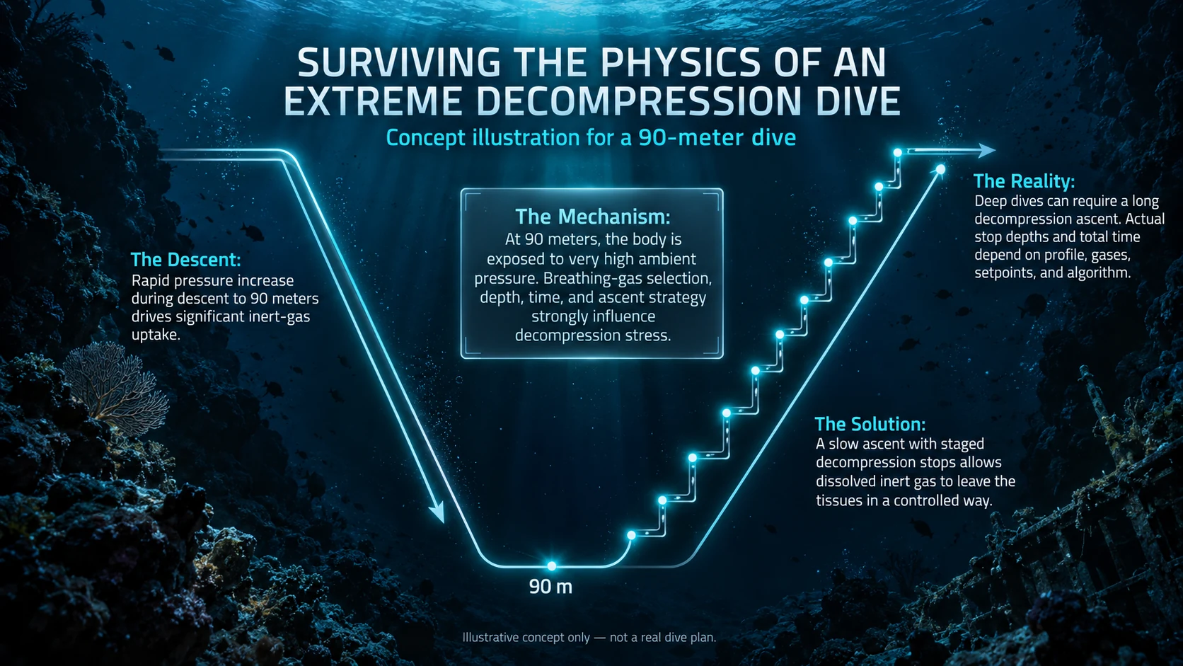
During decompression, dissolved inert gases such as nitrogen and helium leave the diver's tissues as surrounding pressure decreases. However, decompression is not only about dissolved gas: microscopic gas bubbles may also form or grow in tissues and the bloodstream. The relationship between microbubbles, supersaturation and decompression-stop depth is an important topic of continuing research.
1. What Are Microbubbles?
Microbubbles are tiny gas bubbles that may exist or develop during decompression. Their formation and growth are influenced by dissolved inert-gas tension, ambient pressure and physiological factors. Doppler ultrasound can detect small circulating gas bubbles even in divers who experience no symptoms. Excessive bubble formation or growth can contribute to decompression sickness (DCS) by affecting circulation, damaging tissues and promoting inflammation. Not every detected bubble causes injury, and bubble counts cannot directly predict an individual diver's DCS risk.
2. What Is a Deep Stop?
A deep stop is a pause during ascent at a greater depth than would normally be required by a conventional dissolved-gas decompression schedule. The idea became popular through technical diver and marine biologist Richard Pyle, whose observations of post-dive fatigue inspired what became known as Pyle stops, often placed near half of the maximum depth. The hypothesis was that remaining at higher ambient pressure could limit early expansion of existing gas bubbles. However, that is only one part of decompression physiology.
3. The Trade-Off — Bubble Control Versus Inert-Gas Elimination
At deeper stops, ambient pressure is higher. This may limit bubble expansion and supersaturation in fast compartments, but it can also reduce the gradient driving inert-gas elimination. Some slower tissues may continue on-gassing during a prolonged deep stop even while faster compartments are off-gassing. At shallower depths, reduced inspired inert-gas pressure generally increases the opportunity for off-gassing. Extra time deep is therefore not automatically beneficial, particularly if it replaces shallow-stop time.
4. Deep Stops in Decompression Models
Bühlmann ZH-L16 models dissolved inert gas in theoretical tissue compartments. With Gradient Factors, GF Low can influence the initial decompression ceiling and first-stop depth, while GF High influences the permitted gradient near the surface. VPM and RGBM incorporate additional assumptions about microscopic bubble behavior and have historically favored deeper stops in some profiles. A model producing deeper stops is not necessarily safer than one distributing more decompression time at shallower depths.
5. What Does Scientific Research Tell Us?
Some early studies found promising reductions in Doppler-detected bubbles with particular deep-stop approaches. Later evidence complicated that picture. In a major US Navy Experimental Diving Unit (NEDU) study, a deep-stop-heavy schedule produced more decompression sickness than a shallower-stop schedule under the tested conditions. Other experiments have reported increased bubble measurements when shallow decompression time was shifted deeper. This does not mean every deeper pause is harmful; outcomes depend on the entire depth-time profile, gases and distribution of stops.
6. Practical Use for Recreational and Technical Divers
For ordinary recreational NDL diving, there is insufficient evidence to recommend routinely adding deep stops. A controlled ascent and an appropriate shallow safety stop remain established practice. For technical decompression, follow a properly planned and validated schedule: arbitrarily adding deep stops or transferring shallow-stop time deeper may increase decompression stress. Likewise, a very low GF Low should not automatically be interpreted as increased physiological safety.
Recognizing risk and responding appropriately
Missed Decompression — Risks, Symptoms and Emergency Response
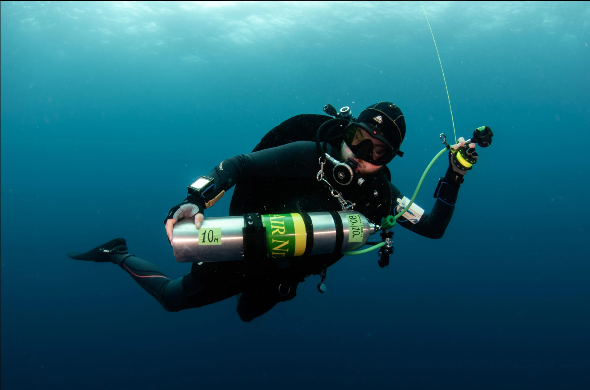
During a decompression dive, required stops allow additional time for inert-gas elimination. If a diver misses a mandatory stop, ascends above the decompression ceiling or surfaces before completing required stops, this is called omitted decompression. It may happen because of insufficient gas, equipment problems, buoyancy difficulties or an emergency.
1. What Happens Inside the Body?
As ambient pressure decreases before enough nitrogen or helium has been eliminated, the tissues may develop excessive supersaturation. Gas bubbles can form or grow in tissues and blood, potentially interfering with circulation, damaging tissues and causing inflammatory reactions. The result may be decompression sickness (DCS), commonly called the bends. Missing a stop increases risk, but does not mean DCS is inevitable; the outcome depends on the exposure and individual factors.
2. How Quickly Can Symptoms Appear?
Symptoms can appear during ascent or within minutes, particularly in severe cases. Many cases emerge within the first hour, and most become apparent within approximately 12 hours. Delayed symptoms can occur after 24 hours or longer, sometimes following additional pressure reduction such as flying. Feeling well immediately after surfacing does not rule out later symptoms.
3. What Types of DCS Do We Recognize?
Type I DCS traditionally refers mainly to joint or muscle pain, some skin symptoms and lymphatic involvement. Type II DCS refers to potentially serious neurological, inner-ear or cardiopulmonary involvement: numbness, weakness, paralysis, poor coordination, vertigo, changes in hearing, trouble breathing, confusion or collapse.
A mottled or marbled rash (cutis marmorata) may indicate important systemic involvement and should never be dismissed as a harmless skin symptom. Modern diving medicine also classifies DCS according to affected organs and severity, rather than relying only on Type I and Type II.
4. Omitted Decompression Without Symptoms
Stop diving and seek prompt diving-medical advice, even if the diver initially feels normal. Avoid flying, travel to altitude and additional dives. Inform the dive team, record the depth/time profile, breathing gases, missed stops and surfacing time, and observe for developing symptoms. Emergency oxygen should be available and provided according to the circumstances, equipment and training, especially after significant omitted decompression.
Do not independently return underwater to complete omitted stops. Specialized professional procedures for in-water recompression exist, but are not general recreational-diving first aid and require specific capabilities and supervision.
5. If Symptoms Appear
Treat symptoms after diving as suspected decompression illness (DCI). Activate emergency medical services, provide the highest appropriate concentration of emergency oxygen using trained procedures, keep the diver resting and protected from exertion and temperature extremes, and contact DAN or a diving-medicine specialist. If fully alert and able to swallow safely, the diver may receive suitable non-alcoholic fluids; never force oral fluids in someone confused, vomiting or unconscious.
Urgent medical evaluation is essential. Professional assessment and, when indicated, hyperbaric oxygen treatment may be required. Temporary improvement with oxygen does not remove the need for evaluation.
6. DCS Versus Arterial Gas Embolism
Arterial gas embolism (AGE) may result from lung overexpansion injury, allowing gas into the arterial circulation. It can cause sudden stroke-like symptoms or unconsciousness during ascent or immediately after surfacing. AGE and DCS involve different mechanisms, but both are forms of decompression illness (DCI) requiring urgent medical attention.
How shallow may we ascend?
Understanding the Decompression Ceiling
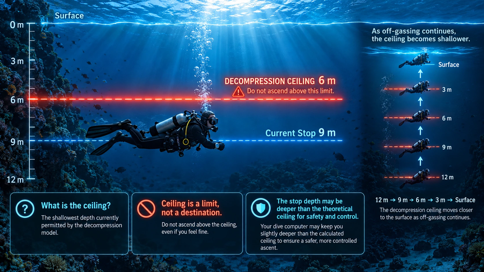
During a decompression dive, the computer continuously estimates inert-gas loading in the diver's theoretical tissue compartments. As ambient pressure decreases during ascent, it calculates how shallow the diver may ascend without exceeding the model's current supersaturation limits. This boundary is called the decompression ceiling.
What Is a Decompression Ceiling?
The ceiling is the shallowest depth currently permitted by the decompression model. If the ceiling is 6 metres, ascending shallower than 6 metres would exceed the model's selected limit. The diver must remain at or deeper than that boundary until the ceiling becomes shallower. A ceiling at 6 metres does not mean the diver must descend or ascend immediately to exactly 6 metres.
How Does the Ceiling Change?
During decompression, dissolved nitrogen and helium are progressively eliminated. As the controlling modeled compartment unloads, the computer recalculates the ceiling. It might move from 12 m → 9 m → 6 m → 3 m → Surface, depending on the profile, breathing gases and decompression settings. Some computers show a continuous ceiling; others display stops in fixed increments.
Ceiling Versus Decompression Stop
A ceiling is the calculated ascent boundary. A decompression stop is the instructed depth where the diver remains to complete decompression. For example, a theoretical ceiling of 4.7 metres may be paired with an instructed stop at 6 metres. Follow the required stop instructions; do not treat the ceiling itself as a target.
What Happens if We Cross the Ceiling?
Ascending shallower than the ceiling is a ceiling violation. At that pressure, modeled tissue loading exceeds the selected supersaturation limit, increasing decompression stress and potentially DCS risk. This is not an exact biological injury threshold, but it must be respected. A computer may warn, recalculate or trigger missed-stop procedures. If a violation occurs, avoid further uncontrolled ascent and follow appropriate computer and emergency procedures when safely possible; never attempt an unsafe return to depth.
Practical Use During Decompression
Monitor the ceiling, required stop depth, ascent rate and remaining obligation; maintain stable buoyancy; and verify that sufficient breathing gas remains. In open water, surge and waves can complicate shallow stops, so trained divers may use a decompression line or other suitable reference.
Constant oxygen partial pressure
Decompression with Constant Oxygen Partial Pressure — CCR and eCCR Diving
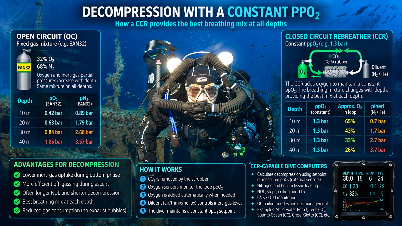
In conventional open-circuit (OC) diving, the diver breathes a fixed gas mixture, so the oxygen and inert-gas partial pressures change with depth. In closed-circuit rebreather (CCR) diving, the breathing gas is recycled and its oxygen partial pressure (ppO₂) can be maintained near a selected setpoint. The gas fractions in the loop therefore change with depth.
How an electronic rebreather works
An eCCR uses a carbon dioxide scrubber to remove CO₂ from exhaled gas. Oxygen sensors monitor loop ppO₂, and an electronic controller can add oxygen through a solenoid to maintain the setpoint. A separate diluent supply provides loop volume and may contain nitrogen, helium or both. The diver must monitor oxygen readings, scrubber limitations and the system's function.
Constant ppO₂ — changing oxygen fraction
For example, at a 1.3 bar ppO₂ setpoint, the idealized oxygen fraction is approximately 65% at 10 m, 43% at 20 m, 32.5% at 30 m and 26% at 40 m. These simple fractions use rounded ambient pressures; actual loop conditions and gas dynamics add complexity. Near the surface, the selected setpoint may no longer be physically achievable.
Why decompression may change
Maintaining an appropriate oxygen partial pressure reduces the pressure available to inert gases compared with many fixed open-circuit mixtures. This can reduce nitrogen or helium uptake, allow longer calculated NDLs, and sometimes shorten decompression. The outcome still depends on depth, time, actual ppO₂, diluent, gas changes and algorithm settings. A higher setpoint also increases oxygen-exposure concerns; it is not automatically the best or safest choice.
Helium and deep CCR diving
For deeper dives, the diluent may be trimix or heliox, helping reduce nitrogen narcosis and breathing-gas density while managing oxygen exposure. Helium is still an inert gas and must be accounted for in tissue-loading calculations. A CCR changes the breathing-gas conditions; it does not remove decompression obligations.
CCR-capable dive computers
A CCR-capable dive computer calculates tissue loading, NDL, ceiling, stops and time to surface (TTS) using a selected or measured ppO₂ and the configured breathing gases. In internal setpoint mode, it assumes the entered ppO₂ has been maintained. In externally monitored mode, a compatible computer can use connected oxygen-sensor readings. Not every CCR computer measures the actual loop ppO₂; a displayed setpoint alone does not prove that the loop reached it.
Open-circuit bailout
If the diver has to leave the breathing loop and switch to open-circuit bailout, the constant-ppO₂ advantage is lost. The computer must use the actual bailout gas and operating mode, and decompression requirements may increase. Suitable bailout gas and a contingency decompression plan must be available for credible failures.
Practical no-decompression dive planning
How to Extend Your Dive Time — Multilevel Diving and Nitrox
For recreational divers, two effective ways to enjoy more underwater time while respecting the No-Decompression Limit (NDL) are multilevel diving and using an appropriate Enriched Air Nitrox mixture.
1. Multilevel Diving — Start Deep, Finish Shallow
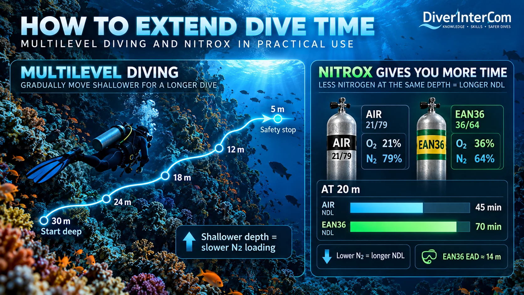
Imagine exploring a reef wall while breathing air. Begin at the deepest planned depth, and then gradually move shallower well before the remaining NDL expires. At the new, shallower depth, inspired nitrogen pressure is lower: nitrogen uptake slows, and some fast compartments may begin off-gassing.
A correctly configured dive computer continuously recalculates tissue loading from the entire depth-and-time profile. The displayed NDL may increase as the diver moves shallower, allowing more total underwater time than remaining at the deepest depth throughout the dive. This is multilevel diving, particularly suited to sloping reefs and reef walls.
Important: A rising NDL does not mean previously absorbed nitrogen has disappeared. Do not repeatedly approach the NDL boundary to extend the dive; leave a sensible margin, monitor gas reserves and finish with a controlled ascent and appropriate safety stop.
2. Nitrox — Less Nitrogen, Longer NDL
Air contains approximately 21% oxygen and 79% nitrogen. Enriched Air Nitrox, such as EAN32 or EAN36, contains more oxygen and less nitrogen. At the same depth, lower inspired nitrogen partial pressure generally means slower nitrogen loading and a longer calculated NDL than on air.
This can also be expressed as Equivalent Air Depth (EAD): the depth at which air would have approximately the same inspired nitrogen partial pressure. At 20 metres on EAN36, the EAD is approximately 14 metres. The diver remains physically at 20 metres, but the nitrogen exposure is comparable to air at about 14 metres.
Nitrox has an essential limitation: higher oxygen partial pressure at depth. The breathing gas must be analyzed and correctly set in the computer, and its Maximum Operating Depth (MOD) and oxygen exposure limits must be respected.
3. Combining Both Methods
A suitable nitrox mix and a progressively shallower profile can work together to reduce unnecessary nitrogen loading. But longer NDL does not mean unlimited dive time: cylinder capacity, breathing-gas reserves, oxygen exposure, conditions and the overall plan still determine when to surface.
Beyond recreational no-stop diving
Extended Decompression — Entering the Realm of Technical Diving
Beyond the No-Decompression Limit
As recreational divers gain experience, they may wish to explore deeper environments or remain underwater longer than conventional no-decompression limits allow. With appropriate advanced training, equipment and experience, the next step is planned decompression diving: dives that deliberately require mandatory decompression stops during ascent.
Short, carefully planned decompression dives can introduce this new discipline. Even a small decompression obligation changes the dive significantly: the diver can no longer ascend directly to the surface without violating the required decompression schedule.
1. Multiple Breathing Gases — More Efficient Decompression
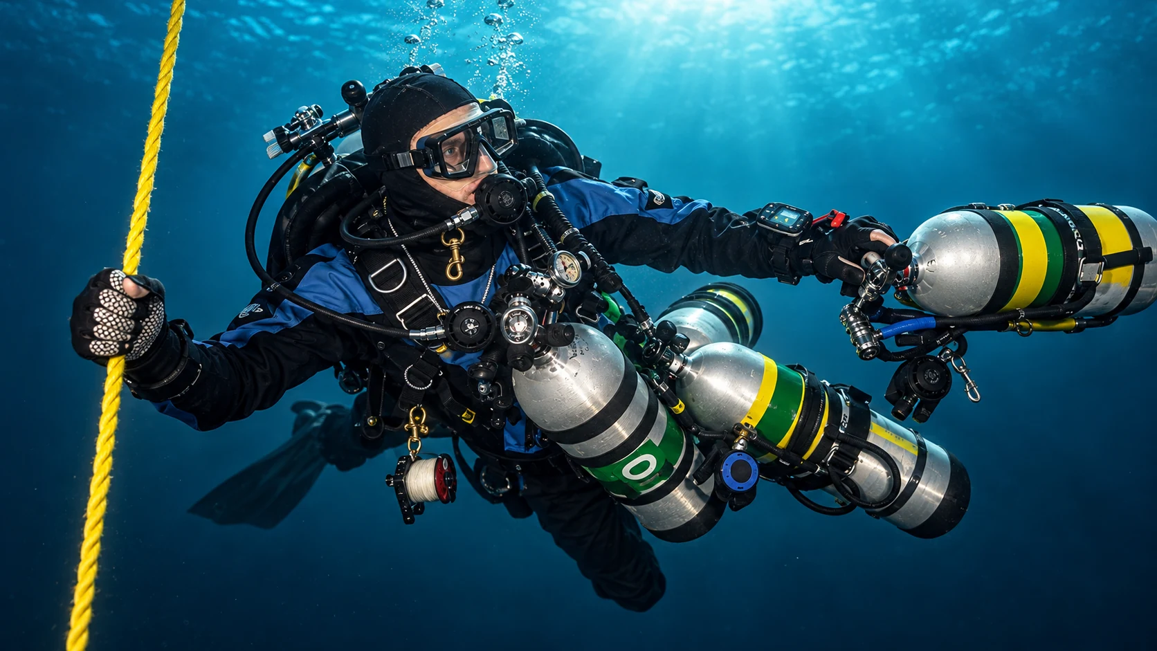
Technical divers may select a bottom gas such as air, nitrox or trimix for the planned depth, and carry additional oxygen-rich gases for decompression. EAN50 (50% oxygen) and EAN80 (80% oxygen) are examples of decompression gases used within their appropriate depth and oxygen-exposure limits.
Switching to a suitable higher-oxygen gas reduces the inspired pressure of nitrogen and helium and promotes inert-gas elimination, potentially shortening the calculated decompression schedule. Each gas has its own maximum operating depth (MOD); gas identification, analysis, switch-depth verification and practice are essential.
2. New Knowledge, Equipment and Responsibilities
Technical decompression diving is more than following additional stops on a computer. Divers need to understand decompression models, oxygen exposure, gradient factors, gas density and consumption, and emergency procedures. Gas management becomes critical: the team must have enough usable gas to complete the dive and decompression, including credible failures or loss of a decompression gas.
Additional cylinders, regulators, redundant systems, reliable buoyancy and precise ascent control require dedicated training and regular practice.
3. Advanced Dive Computers and Planning Software
Technical dive computers can track decompression stops, ceilings, time to surface (TTS), multiple gases and oxygen exposure. For more complex dives, planning programs such as Subsurface, MultiDeco and V-Planner help compare planned profiles, gas changes and decompression settings, while allowing divers to evaluate contingencies and gas requirements.
These calculations depend on the model, settings, correct gas information and realistic execution. A software-generated schedule is not a substitute for training, team planning and adequate reserves.
4. Explore Decompression with DiverInterCom KCore
Try our DiverInterCom Decompression Simulators to explore how dive profiles, breathing gases and settings change theoretical decompression outcomes. These educational and experimental tools are not validated operational dive planners and must not be used to conduct real decompression dives.

Inside the mathematics of dive planning
Advanced Decompression Theory
Understanding the Science Behind Advanced Decompression
As divers progress into technical diving, decompression becomes more than simply following the stops displayed on a dive computer. Advanced divers benefit from understanding how decompression models calculate inert-gas loading, why different tissues control the ascent, and how nitrogen, helium, breathing-gas composition and gradient factors affect decompression obligations.
Modern dive computers use mathematical models to estimate these processes. One of the most widely used is Bühlmann ZH-L16, often combined with adjustable gradient factors. Understanding the mathematical foundations helps divers interpret their computers, evaluate dive plans and recognize the limitations of decompression calculations.
1. Tissue Compartments and Half-Times
The Bühlmann ZH-L16 model represents the body using 16 theoretical tissue compartments. These compartments are mathematical representations of different rates of inert-gas uptake and elimination. Each compartment has a characteristic half-time, describing how quickly its inert-gas pressure approaches equilibrium with the inspired inert-gas pressure. Fast compartments respond rapidly to pressure changes, while slow compartments require much longer to approach equilibrium.
2. Haldane's Equation — Gas Loading at Constant Depth
At a constant depth with constant inspired inert-gas pressure, tissue loading follows an exponential function. This explains why a fast compartment approaches equilibrium quickly while a slow compartment may continue absorbing inert gas throughout a long dive, and why decompression obligations depend on both depth and exposure time.
Pₜ(t) = Pᵢ + (Pₜ₀ − Pᵢ) × e^(−kt)
k = ln(2) / T½
Pₜ(t): tissue inert-gas pressure after time t; Pₜ₀: initial tissue pressure; Pᵢ: constant inspired inert-gas pressure; k: rate constant; T½: compartment half-time.
3. The Schreiner Equation — Gas Loading During Ascent and Descent
Real dives rarely remain entirely at one depth. During ascent and descent, inspired inert-gas pressure changes continuously. The Schreiner equation extends the basic tissue-loading model by describing how tissue gas changes when inspired pressure changes linearly with time. Dive-planning software may use Schreiner calculations or numerical integration to model changing-depth profiles.
Pₜ(t) = Pᵢ₀ + R(t − 1/k) − (Pᵢ₀ − Pₜ₀ − R/k) × e^(−kt)
Pᵢ₀: initial inspired inert-gas pressure; Pₜ₀: initial tissue pressure; R: rate of change of inspired inert-gas pressure; k: compartment rate constant; t: elapsed time. Apply separately to each inert gas.
4. Nitrogen and Helium — Two Different Inert Gases
When breathing trimix, both nitrogen and helium contribute to inert-gas loading. Bühlmann models these gases separately because they have different half-times and decompression coefficients. The total inert-gas pressure in a compartment is the combined effect of both gases. Helium generally has faster modeled tissue kinetics than nitrogen, but that does not automatically mean shorter decompression. The outcome depends on the full dive profile, gas mixture and decompression settings.
5. Bühlmann M-Values — The Mathematical Ascent Limit
Bühlmann's decompression model defines a relationship between ambient pressure and the maximum tolerated tissue inert-gas pressure. These model limits are called M-values. They are mathematical thresholds, not guaranteed biological boundaries between safe and unsafe decompression.
M = a + Pₐₘᵦ / b
M: modeled tolerated tissue inert-gas pressure; Pₐₘᵦ: ambient absolute pressure; a and b: compartment-specific coefficients (combined appropriately for nitrogen–helium mixtures).
6. The Leading Tissue — Which Compartment Controls Decompression?
At any moment, the 16 compartments can have different inert-gas pressures and different decompression limits. The leading tissue, also called the controlling compartment, is the compartment currently imposing the most restrictive decompression ceiling. It is not necessarily the fastest tissue or the one containing the greatest amount of inert gas. It is simply the compartment closest to its allowed limit under the chosen model and settings, and it may change several times during a single dive.
7. Gradient Factors — Adding Conservatism to the Model
Gradient factors modify the allowable supersaturation relative to Bühlmann's modeled M-values. GF Low mainly influences the beginning of the decompression profile, while GF High defines the selected limit near the surface. Lower settings usually mean more conservative calculated decompression, but they are mathematical adjustments to the model, not direct percentages of decompression-sickness probability.
Pₐₗₗₒwₑd = Pₐₘᵦ + GF × (M − Pₐₘᵦ)
GF is a decimal fraction (for example 0.85 for 85%). M is the Bühlmann limit at that ambient pressure. The schedule varies GF along the ascent according to the selected GF Low and GF High settings.
8. Gas Density — The Physiological Side of Technical Diving
Decompression calculations are only one part of advanced dive planning. Another important factor is breathing-gas density, which increases with ambient pressure. Dense breathing gas increases breathing resistance and may contribute to carbon dioxide retention. Helium is much less dense than nitrogen, so replacing some nitrogen with helium can reduce breathing-gas density at depth. Gas density is not itself a Bühlmann decompression variable, but it is an important practical part of advanced gas planning.
9. Beyond the Mathematics — The Limits of Decompression Models
Modern decompression models are powerful planning tools, but they remain simplified representations of complex human physiology. Real decompression is influenced by factors such as circulation, microbubbles, exercise, temperature, hydration and previous dives. The mathematical compartments are not individual organs, and modeled inert-gas pressure is not a direct measurement of a diver's actual tissues.
10. Applying Advanced Decompression Theory
Understanding these principles allows experienced divers to interpret advanced dive-computer information more meaningfully. With suitable planning software, divers can explore how depth, bottom time, gases, the leading tissue and gradient factors affect calculated decompression. Examples of established planning tools include Subsurface, MultiDeco and V-Planner. The DiverInterCom KCore decompression simulators can also be used to explore these mathematical principles in an educational environment.
Want to know more? Stay tuned. Dive in. Explore further.
Use KCoreCopilot to explore Decompression Theory